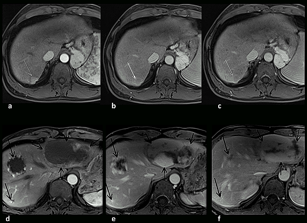
Figure 3:
A cirrhotic patient evaluated for possible HCC (a – c) and a patient evaluated for indeterminate liver mass (d – f). The hepatic arterial dominant phase (HADP) (a, d), early hepatic venous phase (EHVP) (b, e) and interstitial phase (c, f) were observed on transverse 3D-GE images acquired on 3.0 T. The first patient displayed a lesion (white arrow a - c) with central diffuse enhancement on HADP, with rapid washout and peripheral capsular enhancement developing on EHVP and interstitial phase (corresponding to a pseudocapsule). These MR imaging features are characteristic of HCC. Prominent varices are also observed (thin black arrow a – c). The second patient displayed three lesions (black arrows d – f) with discontinuous peripheral nodular enhancement on HADP and progressive centripetal enhancement on subsequent phases. These MR imaging features are characteristic of hemangioma. The greatest lesion occupies a large portion of left lobe and shows persistence of a central scar on interstitial phase imaging, consistent with a type 3 hemangioma.
|