|
||||||||||||||||||||||||||||||||||||||
|
|
||||||||||||||||||||||||||||||||||||||
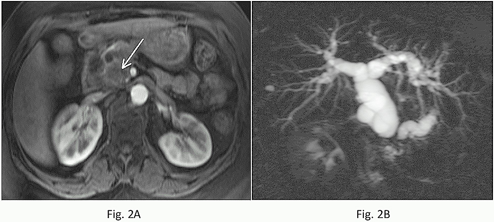
Fig.: 2A-2B:
Pancreatic adenocarcinoma arising in the head. Transverse immediate post-contrast fat-suppressed T1-weighted 3D-GE (A) and coronal oblique thick-section MRCP (B) images. On immediate postcontrast image (A), the tumor is well shown as a low signal intensity mass (arrow). The MRCP image (B) demonstrates obstruction of the common bile duct and pancreatic duct, creating the double duct sign.
|
||||||||||||||||||||||||||||||||||||||