|
||||||||||||||||||||||||||||||||||||||
|
|
||||||||||||||||||||||||||||||||||||||
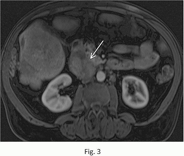
Fig.: 3:
Functioning islet cell tumor. T1-weighted post-contrast fat-suppressed 3D GE image shows diffuse heterogeneous enhancement of the tumor (arrow).
|
||||||||||||||||||||||||||||||||||||||
|
||||||||||||||||||||||||||||||||||||||
|
|
||||||||||||||||||||||||||||||||||||||
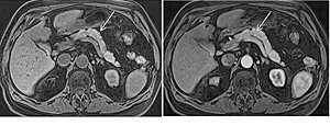
Fig.: 3:
Functioning islet cell tumor. T1-weighted post-contrast fat-suppressed 3D GE image shows diffuse heterogeneous enhancement of the tumor (arrow).
|
||||||||||||||||||||||||||||||||||||||
![]()